Pet Corneal Mineral Dystrophy in Katy, TX
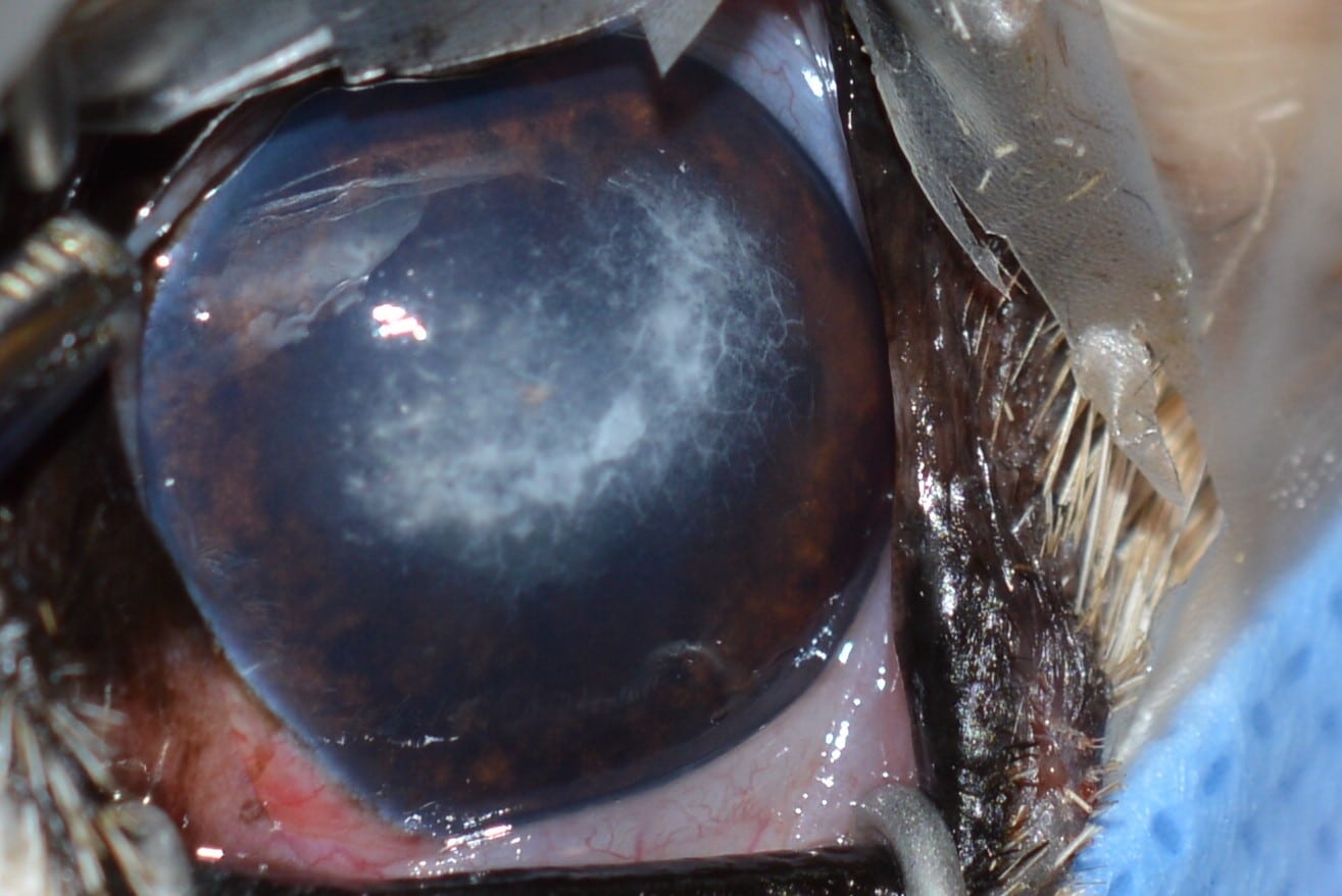
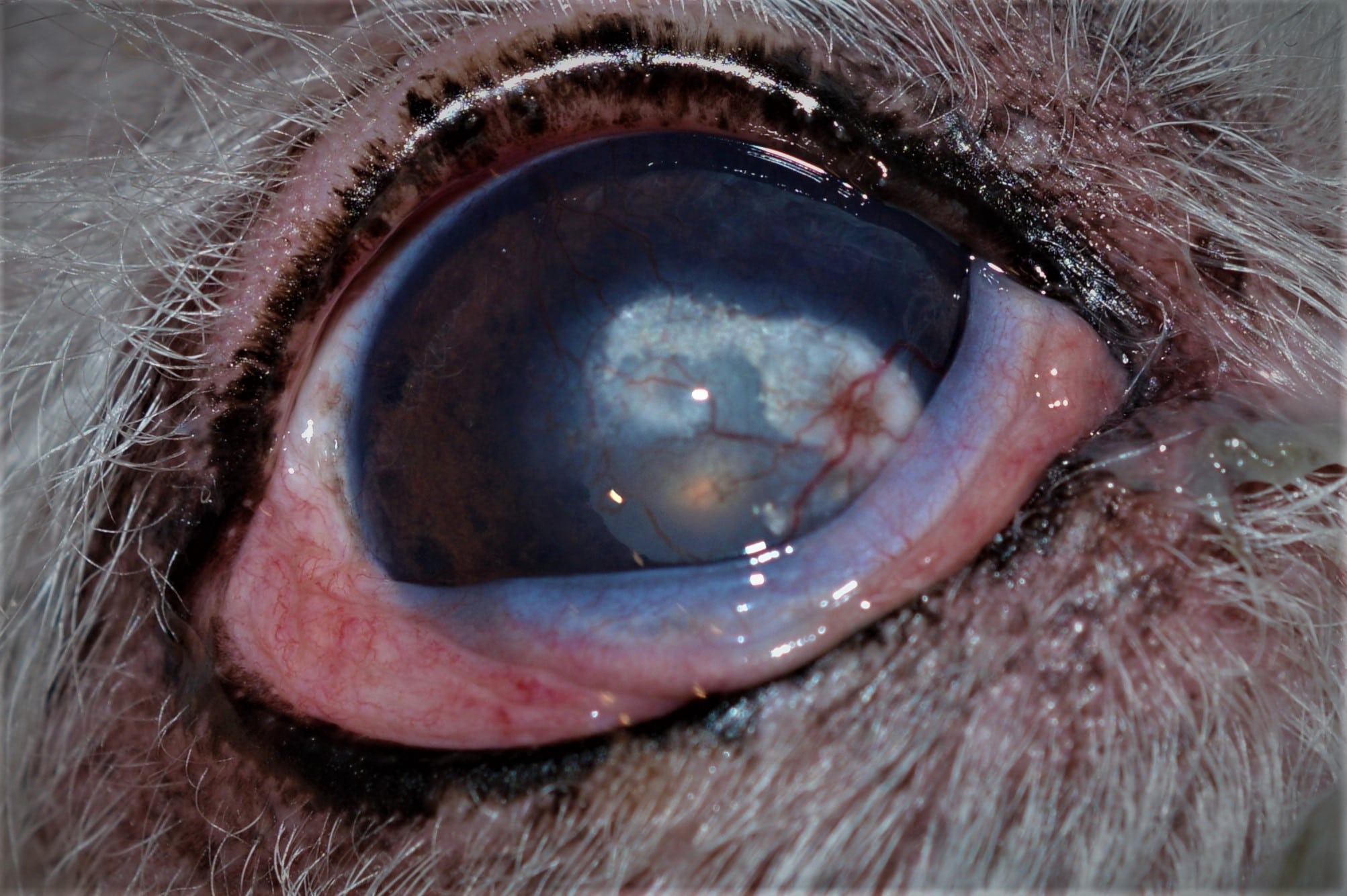
Corneal mineral dystrophy is a condition that affects pets’ eyes, leading to the formation of white, opaque spots on the cornea. These spots are a result of mineral deposits that accumulate in the corneal tissue. At Animal Eye Medical & Surgical Specialists in Katy, TX, we provide specialized care for pets with corneal mineral dystrophy, ensuring their eye health is carefully monitored and managed.
What is Corneal Mineral Dystrophy?
Corneal mineral dystrophy is characterized by the buildup of mineral deposits, such as calcium, within the corneal tissue. These deposits appear as white, circular lesions, typically in the central part of the cornea. The condition is often bilateral, meaning it can affect both eyes and usually remains stable without significant progression. While corneal mineral dystrophy generally does not cause pain or significant vision loss, monitoring the condition to prevent potential complications is important.
Symptoms of Corneal Mineral Dystrophy
Recognizing the signs of corneal mineral dystrophy early can help in managing the condition effectively. The symptoms are usually mild but can include:
- White or grayish spots on the cornea
- Lesions that are typically circular and located in the center of the eye
- Minimal to no vision impairment
- Lack of associated pain or discomfort
These symptoms are often more noticeable in older pets, although the condition can develop at any age. Regular eye examinations are essential to ensure that the condition does not worsen or lead to other complications.If you notice any of these symptoms in your pet, contact us immediately. Early intervention can prevent further issues and ensure your pet’s eye health is maintained.

Causes of Corneal Mineral Dystrophy
The exact cause of corneal mineral dystrophy is related to the metabolism of the corneal cells. It can occur as a primary condition or secondary to other underlying health issues. Some common causes include:

- Aging: Changes in the metabolism of corneal cells as pets age can lead to mineral deposits.
- Chronic Corneal Ulceration: Persistent corneal ulcers can contribute to mineral deposition.
- Hormonal Imbalances: Conditions such as Cushing’s disease can affect corneal health.
- Chronic Use of Steroid Eye Drops: Prolonged use of certain medications can lead to corneal changes that result in mineral deposits.
Identifying and managing any underlying conditions is crucial in preventing the progression of corneal mineral dystrophy.
Treatment Options for Corneal Mineral Dystrophy
In most cases, corneal mineral dystrophy does not require treatment, especially if the condition is mild and does not cause discomfort or vision loss. However, when treatment is necessary, the following options may be considered:
Regular veterinary check-ups to monitor the condition and ensure it remains stable.
Eye drops may be prescribed to manage mineral deposition and prevent progression.
In severe cases where vision is affected, or the condition causes discomfort, a surgical procedure called keratectomy may be performed to remove the affected corneal tissue.
Why Choose Animal Eye Medical & Surgical Specialists?
- Specialized care for complex eye conditions
- Advanced diagnostic and treatment techniques
- Personalized attention and follow-up to ensure the best outcomes
- Compassionate approach to managing your pet’s eye health
If you suspect your pet may have corneal mineral dystrophy or degeneration, contact Animal Eye Medical & Surgical Specialists in Katy, TX, right away. Our experienced team is ready to provide the expert care your pet needs to manage this condition effectively. Call us today at 832-437-0119 to schedule an appointment and learn more about how we can help maintain your pet’s eye health.
