Pet Retrobulbar Masses in Katy, TX
Pet retrobulbar masses are serious conditions that affect the area behind the eye, often leading to discomfort, pain, and potential vision loss. Recognizing the signs early and seeking prompt veterinary care is crucial for managing this condition effectively. At Animal Eye Medical & Surgical Specialists in Katy, TX, we provide expert diagnosis and treatment for pets with retrobulbar masses.
What Are Pet Retrobulbar Masses?
A retrobulbar mass refers to a growth or lesion located within the orbit (eye socket) behind the eye. These masses can be caused by various conditions, including infections, cysts, or tumors, and can have different impacts depending on their nature and location.
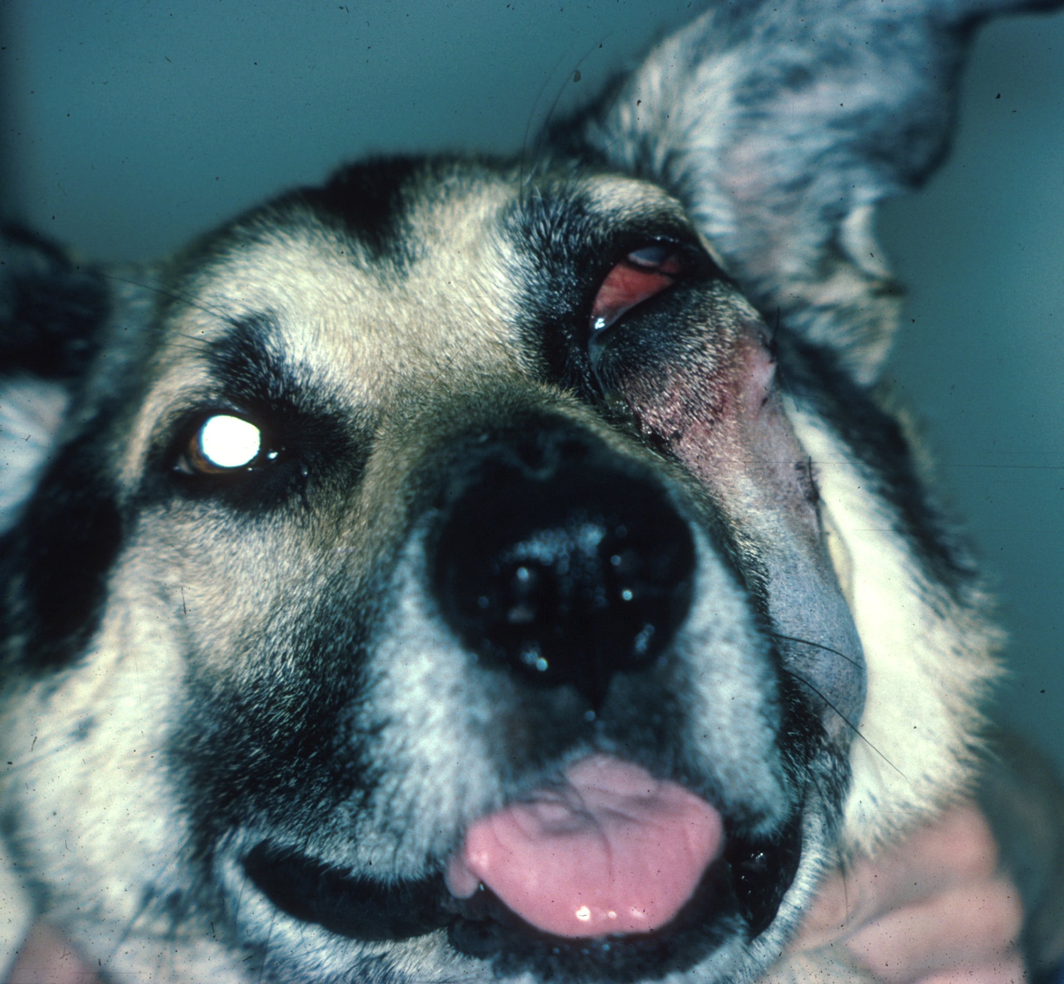
Pets with retrobulbar masses often display several clinical signs, which may vary based on the underlying cause and the mass’s severity. Common symptoms include:
- Protrusion of the Eye (Exophthalmia): The affected eye may bulge outward due to the mass pushing the globe forward.
- Third Eyelid Protrusion: The third eyelid may become more visible as it moves forward over the eye.
- Redness and Swelling: Inflammation around the eye and surrounding tissues can lead to noticeable redness and swelling.
- Pain and Discomfort: Pets may avoid being touched near the eye, have difficulty opening their jaws, and show signs of pain such as vocalization or lethargy.
- Periorbital Swelling and Discharge: Swelling around the eye socket and abnormal discharge can be present.
- Vision Loss: Depending on the size and location of the mass, blindness in the affected eye may occur.
If you notice any of these symptoms in your pet, prompt evaluation is essential. Contact us today to schedule an appointment and ensure your pet receives the specialized care they need to protect their vision and comfort.

Causes of Retrobulbar Masses
The development of retrobulbar masses can result from various conditions, including:
Infections that lead to the formation of pus-filled abscesses or inflammation of the tissues can cause rapid swelling and pain. This is commonly seen in younger, large-breed dogs.
A cystic structure of the salivary gland, often developing slowly and painlessly, is more common in breeds like Boston Terriers.
Tumors can develop within the orbit or spread from other areas of the body. In dogs, these tumors tend to be benign, while in cats, they are more likely to be malignant.

Diagnosing Retrobulbar Masses
Accurate diagnosis of retrobulbar masses is essential for effective treatment. Diagnostic procedures may include:
- Ocular Ultrasound: This imaging technique can help detect the presence of a mass and guide sample collection for further testing.
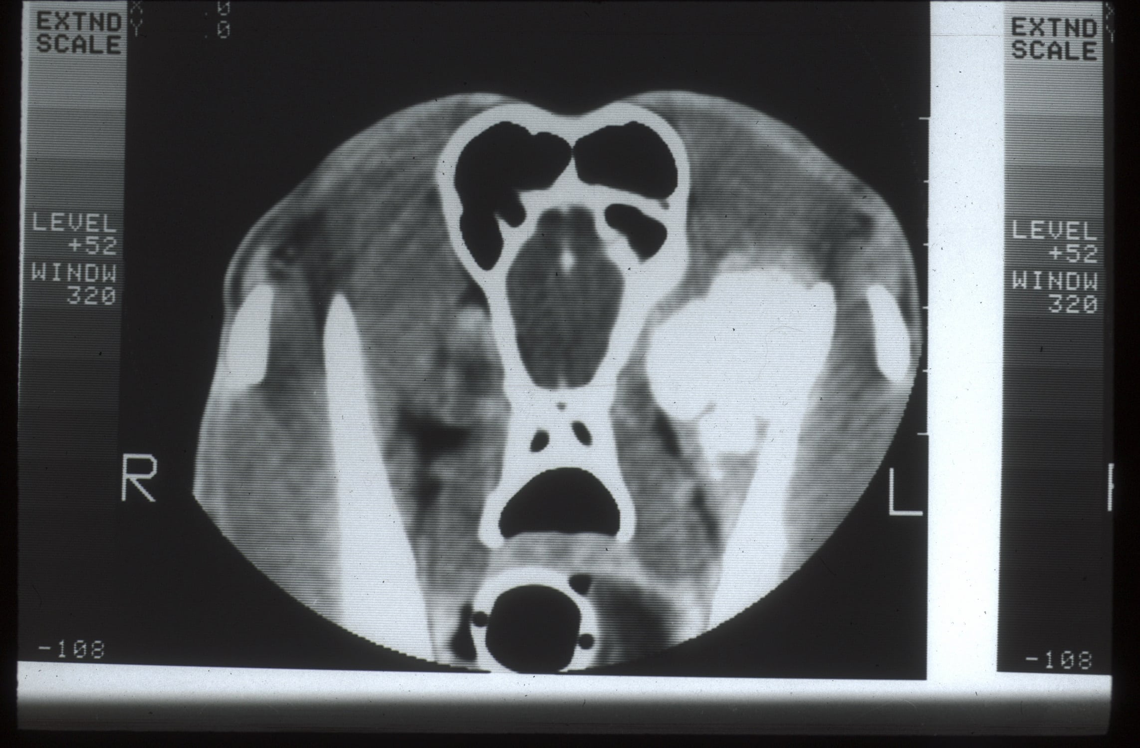
- Advanced Imaging (CT Scan or MRI): These methods provide a detailed view of the orbit, helping to determine the extent of the mass, its origin, and whether it has spread to surrounding tissues.
Treatment Options for Retrobulbar Masses
The treatment for retrobulbar masses depends on the type of mass, its location, and the overall health of the eye. Treatment options include:
Abscess/Cellulitis Treatment
Treatment typically involves creating a draining tract from the root of the mouth to the orbit under anesthesia, along with systemic antibiotics and anti-inflammatory medications.
Sialocele Management
Management may involve periodic drainage, the injection of a sclerotic agent to promote scarring, or surgical removal of the cyst.
Cancer Treatment
Depending on the tumor’s severity, surgical removal may be required, followed by chemotherapy or radiation therapy. In some cases, the entire orbit, including the eye, may need to be removed if the cancer is extensive.
Key Services at Animal Eye Medical & Surgical Specialists
- Comprehensive Eye Examinations: Detailed assessments to detect and diagnose retrobulbar masses and other ocular conditions.
- Advanced Imaging Techniques: Utilizing CT scans, MRIs, and ocular ultrasounds for accurate diagnosis and treatment planning.
- Personalized Treatment Plans: Tailored care based on your pet’s specific needs, ensuring the best possible outcomes.
- Experienced Veterinary Ophthalmologists: Specialized care from a team with extensive expertise in managing complex eye conditions.
At Animal Eye Medical & Surgical Specialists in Katy, TX, we are dedicated to providing expert care for pets with retrobulbar masses. Call us today at 832-437-0119 to schedule a consultation if your pet is showing signs of a retrobulbar mass. Early detection and treatment can make a significant difference in your pet’s health and quality of life.

